Congratulations to the winners of the STS Elionix Scientific Image competition of Q1 2022!
The STS Elionix Scientific Image competition allows us to showcase the creative side of the researchers here at Tyndall. They share scientific images from their research which they have discovered to be visually striking or informative to the technically excellent.
The judges for the Q1 competition were Prof. William Scanlon, CEO; Prof. Eoin O'Reilly, Chief Scientist; Peter Smyth, Commercial Director; Ursula Morrish, Marketing & Communications Manager; Graeme Maxwell, Head of Specialty Products & Services; and Dr Daniela Iacopino, Researcher, MNS.
The exceptional quality and creativity in these images led to the votes being cast to two talented winners: Dr. Emmanouil Amargianitakis and Dr Vuslat Juska.
Congratulations again to the winners.
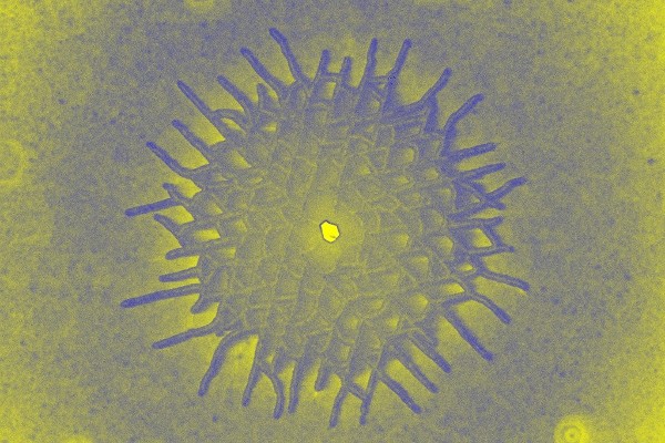
Crystal Starburst
Dr. Emmanouil Amargianitakis, III-V Materials and Devices Group

This image shows the central region of a circular defect which has the shape of a crisscross grown on the surface of a semiconducting crystal. The image was taken with a scanning electron microscope.
The goal is to exquisitely control the properties of charge in semiconductor materials, and hence, to engineer the light emitting properties.
The feature was created unintentionally during the high temperature (1000 degrees Celsius) growth of GaN/AlGaN materials by metal organic chemical vapor deposition. These are the materials used in the now ubiquitous Light Emitting Diodes (LEDs). The purpose of the growth was to create a periodic sequence of ultrathin layers (each 10 atomic planes) of two different materials that can be utilized to precisely confine positive and negative charges for their use in laser applications in the ultraviolet (UV) range.
In the image, the spread of the defect follows a specific direction that is influenced by the hexagonal symmetry of these materials. This work is part of the Irish Photonic Integration Centre (IPIC) project “Realization of ultra-violet III-nitride solid state lasers employing micro-transfer printing for their integration with waveguides” at Tyndall.
NanoButterflies
Dr Vuslat Juska, Nanosensors

Nanobutterfly-arrays-on-a-chip is a silicon based micro fabricated device designed and fabricated during Dr Juska's Catalyst project at Tyndall. This project’s aim is to develop an electrochemical biosensing platform based on silicon nanotechnologies; therefore during this project Dr Juska designed and developed several silicon based chips which provide excellent platforms to achieve varying microarchitectures as interfaces.
As such, the image represents one of the designs dedicated for immunosensor development. The overall chip is a highly miniaturised device composed of multiple sensing electrodes and a pair of shared counter-reference electrodes. The image is taken after the metal electrodeposition. Each sensing area has seven ultramicro scaled recessed electrodes which are arranged at the same distance to the adjacent electrodes. The design is highly critical for such small scale of the arrays to exhibit microelectrode capabilities.
The controlled electrodeposition conditions of gold created butterfly-like nano-architectures on each electrode on the array. The walls of the wings are highly porous therefore these butterflies provide increased surface areas for electrochemical applications, which is one of the most desired conditions at ultra-micro scale. The aim is to transform these nano-architectures into sensing platforms to serve as diagnostic tools for neurodegenerative diseases, in particular Multiple Sclerosis.
NanoIceberg
Dr Vuslat Juska, Nanosensors Group

This SEM image represents one of the nanotextured interfaces on an array that Dr Juska developed for a Catalyst project at Tyndall. The project aims to develop a diagnostic tool for Multiple Sclerosis.
The blue iceberg-like architecture is electrodeposited gold with highly porous walls. The surface morphology is key to overcome the limitations of many electrochemical biosensing systems.
The high porosity of the walls provides increased surface area therefore the overall device is capable of providing enhanced electrical signal. Dr Juska’s goal is to use these depositions as an immobilisation matrix for biorecognition elements of interest in order to develop rapid and sensitive detection tools for neurodegenerative diseases, such as Multiple Sclerosis.