Tyndall extends its congratulations to the winners of the STS Elionix Scientific Image Competition of Q2 2025!
Open to all researchers at Tyndall, the competition provides a platform to celebrate the artistic side of science, as seen through the microscope. Entrants were invited to submit images that not only reflect their research but also captivate the eye.
The judging panel for this quarter included Professor William Scanlon, CEO, Tyndall; Dr Graeme Maxwell, Head of Specialty Products & Services; Dr Daniela Iacopino, Researcher, MNS; Ursula Morrish, Marketing and Communications Manager
The judges selected three winning entries based on their originality, visual impact, and scientific relevance. The winners of the Q2 2025 competition are:
- Aashi Gupta, Pavlina Metaxa, Brendan Sheehan & Ray Duffy – “WS2 Nachos in a salsa spread”
- Atieh Mousavi & Alida Russo – “Fairy Forest”
- Christian Corcoran & Chandrashekar Lakshmi Narayana – “Dancing Crayons”
Each winner will receive a €150 Me2You Gift Card, generously sponsored by STS Elionix, in recognition of their contribution to scientific communication and visual storytelling.
WS2 Nachos in a salsa spread
By Aashi Gupta, Pavlina Metaxa, Brendan Sheehan & Ray Duffy, Quantum Electronic Devices (QED) Group
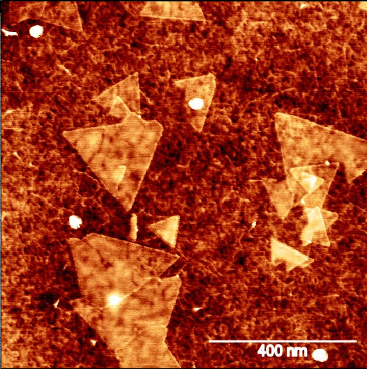
This scientific image is an AFM image of MOCVD grown ~1.2 monolayers of Tungsten Disulphide (WS2).
The ‘salsa dip’ represents the monolayer of tungsten disulphide (WS2) with uniform grain sizes, whereas the triangles (‘nachos’) are the WS2 grains that coalesce to form bigger grains due to the high thermal budget.
This image was taken using the Bruker Dimension Icon AFM tool at the OACF lab in Tyndall using a high-resolution tip. This image demonstrates the capability of the tool to take high-resolution images even for very small scan sizes of around 400nm. This image can further be used to calculate the grain sizes of WS2, which is a crucial parameter to determine the device performance.
.
Fairy Forest
By Atieh Mousavi & Alida Russo, Life Science Interface (LSI) Group
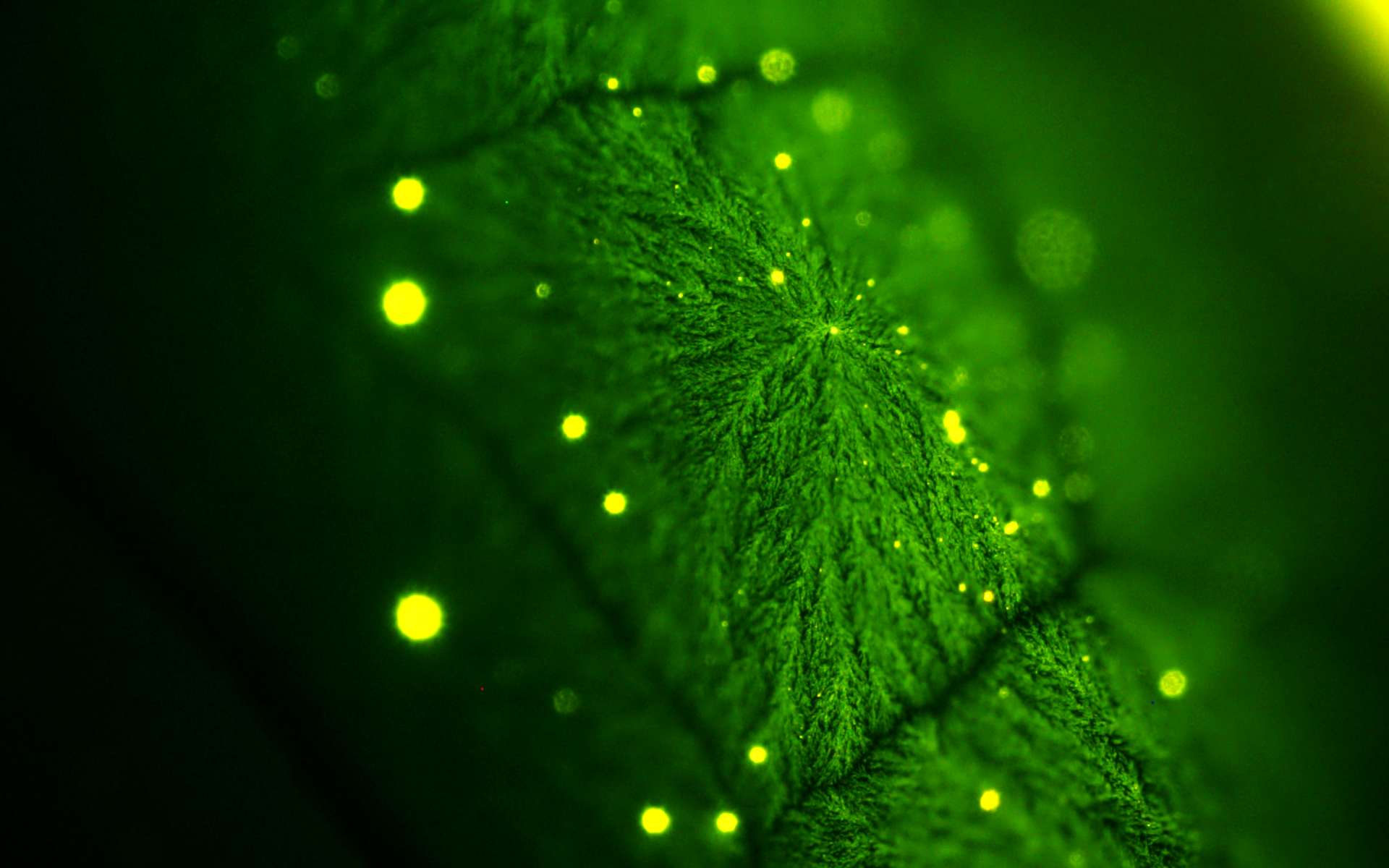
This is a fluorescence image of antibodies on the surface of a polymer modified electrode that resembles Irish Faires in a Forest.
The aim of this project is to develop a disruptive multiplexed near-patient diagnostic solution to enable the early detection of Stage I and II ovarian cancer using different biomarkers. Designing, fabricating, and characterising our sensors to be used as detection tools. This image was taken after the functionalization of homemade electrodes with polymers and antibodies.
The innovative approach focuses on creating highly sensitive and specific sensors that can detect multiple biomarkers simultaneously. The unique fibrous structures of the polymer layer, influenced by the conditions of polymerization, contribute to the improved conductivity and surface area of the electrodes. This allows for better immobilization of antibodies and more effective electrochemical sensing, ultimately aiming to improve patient outcomes through earlier diagnosis and treatment.
Dancing Crayons
By Christian Corcoran & Chandrashekar Lakshmi Narayana, Nanophotonics Group & Quantum Electronic Devices Group
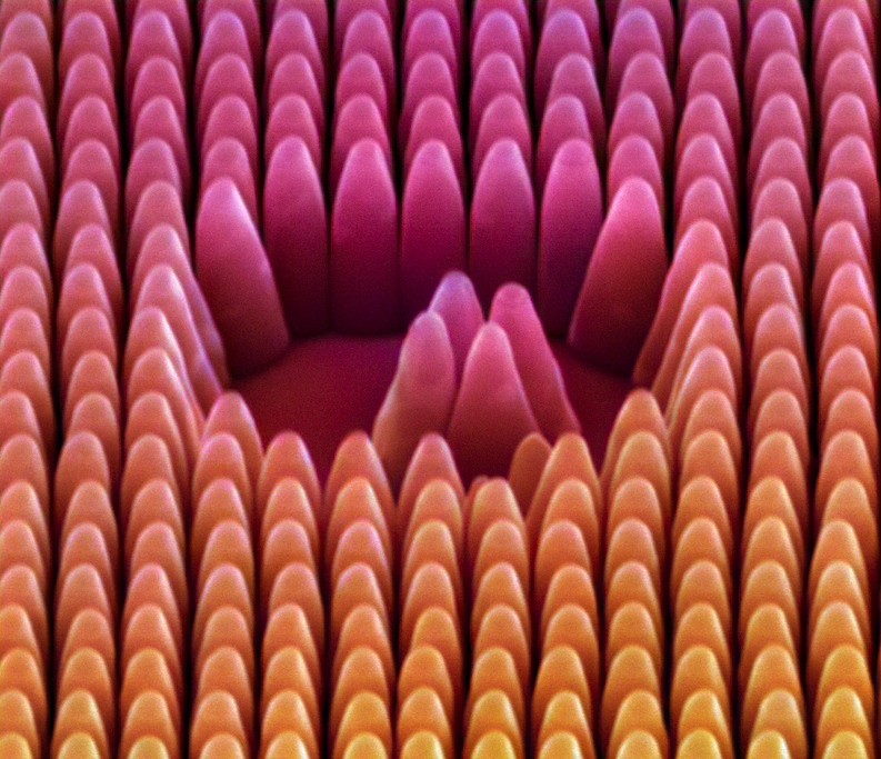
By a wet-etch process these pillars are revealed from their Si substrate.
These metasurfaces can be employed to achieve low PPB concentrations for photothermal spectroscopy. The near-field enhancement effect is beneficial for Mid-IR sensing, especially while working with low sample volumes.
