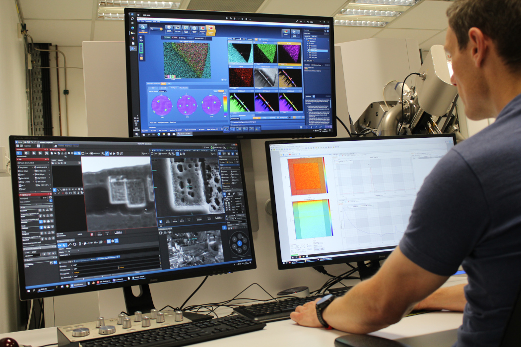
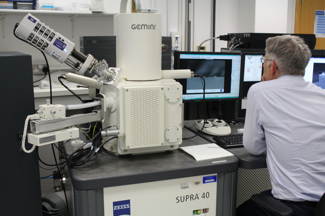
The Electron Microscopy and Analysis Facility (EMAF) delivers state-of-the-art electron microscopy analyses with rapid turnaround time for industry and academia

Electron Microscopy
We develop customer- and product-specific analysis of materials and devices and provide comprehensive understanding of the measurement results:
- Product contamination analysis
- Thin film analysis (thickness + structure)
- Failure analysis
- Materials characterisation
- Surface analysis
- (S)TEM sample preparation
- Trace analysis >10ppm
- 3D visualisation
- Correlative microscopy
- Focused ion beam (FIB) patterning/prototyping.

Methods
- Scanning electron microscopy (SEM)
- Elemental analysis (EDX and ToF-SIMS)
- (Scanning) transmission electron microscopy (S)TEM
- Dual beam focused ion beam (FIB)
- Electron Backscatter Diffraction (EBSD)
- Electron diffraction (SAED)
- Electron column for UHR imaging (immersion lens for non-magnetic samples only)
- Analysis mode: SE detector, for topography contrast
- UHR-resolution mode: In-beam SE
- Backscattered Mode: In-Beam f-BSE, Mid angle BSE
- Retractable BSE detector, for wide angle backscattered electrons for material contrast
- Retractable HADF STEM (R-STEM) detector: Bright field, Dark field and High Angle Dark Field
- SEM Resolution
- 0.6nm @30kV, 20kV, and 15kV
- 0.7nm @10kV
- 1.0nm @5kV
- 1.2nm @1kV
- 1.7nm @500V

Methods
Ga ion FIB column
- Aligned at 55 degrees to Electron column
- For cross sectioning, 2D/3D patterning, 3D tomography and TEM lamella preparation
Xe ion FIB column
- Aligned at 55 degrees to Electron column
- For cross sectioning, 2D/3D patterning, and 3D tomography
Nanoflat Delayering
- Milling rate up to 1000 µm3/s
Detectors
- Oxford Aztec UltiMax 170 EDX & elemental analysis system
- Oxford Symmetry Electron Backscatter Spectroscopy (EBSD)
- Tofwerk Time of Flight Secondary lon Mass Spectroscopy (ToF-SIMS)
Electron column for UHR imaging
- Analysis mode: SE detector, for topography contrast
- UHR-resolution mode: In-beam SE
- Backscattered Mode: In-Beam f-BSE, Mid angle BSE
- Retractable BSE detector, for wide angle backscattered electrons for material contrast
- Retractable HADF STEM (R-STEM) detector: Bright field, Dark field and High Angle Dark Field
3D Virtual Lab Tour